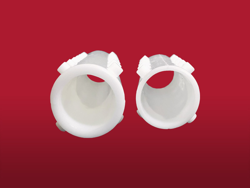
Insertion technique
The procedure is performed under general anesthesia. The implant can be carried out directly through the working channel of the tracheoscope or bronchoscope, or by using a conventional introducer for silicone prostheses. The airway is accessed with a rigid endoscope.
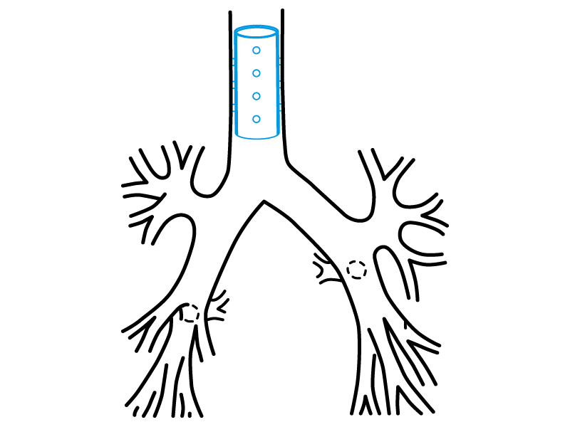
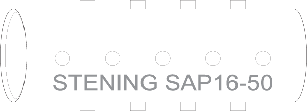
The length and diameter of the area to be covered with the stent must be properly established. A simple method to determine the length of the affected area is to mark the tracheoscope when its tip reaches the end of the lesion, and to repeat the marking after withdrawing it back to the beginning of the lesion. The diameter of the trachea must be estimated by comparison with the known diameter of the endoscope used.
Retrograde implantation method
- Lubricate the mouthpiece of the introducer with lidocaine gel, avoiding contact between the lubricant and the operator’s fingers.
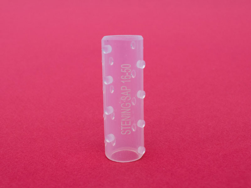
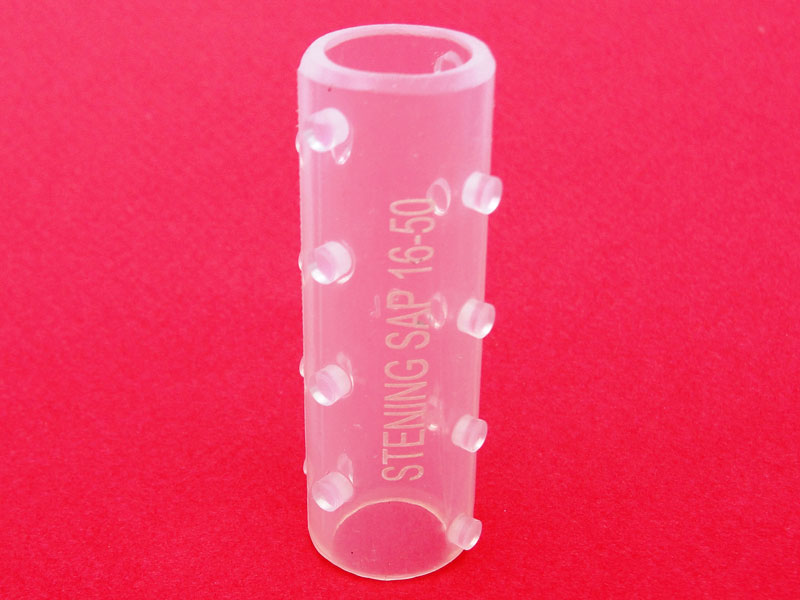
- Fold the Stening® along its axial axis and insert it into the prosthesis introducer through the mouthpiece.
- Remove the mouthpiece.
- Advance the tracheoscope tube beyond the lesioned area and place its distal end or bevel on healthy mucosa, exceeding the affected zone by about 5 to 7 mm.
- Place the introducer inside the tracheoscope.
- Press the ejector while withdrawing the tracheoscope by the same amount the ejector plunger advances: the plunger of the stent loader is pressed as the endoscope is withdrawn.
The prosthesis is thus released. If necessary, it can be repositioned with an alligator forceps, the maneuver being simpler if the stent is placed “below” the lesion.
Antegrade implantation method
Steps 1, 2 and 3 are repeated. Then the tracheoscope containing the introducer and the prosthesis is stopped 5 mm before the lesion to be treated, and the ejector plunger is pressed slowly. In this way, the prosthesis will be expelled toward the affected trachea.
Some stent loader models are not introduced inside the tracheoscope, but simply attached to it at its proximal end, from where the stent is propelled. For this purpose, the endoscope will have been stopped proximally or distally to the lesion as explained above, in order to push the prosthesis with the plunger provided with the endoscopic instrument. The stent will then travel through the entire interior of the tracheoscope until reaching the trachea. At this point, a sudden reduction in resistance to the pressure exerted on the plunger will be perceived, indicating that the stent has begun to exit the interior of the endoscope.
Correction of the stent position
The stent may require additional maneuvers in order to correct or adjust its final position. It is preferable to correct a stent that has been installed beyond the desired position than the opposite, since it is highly inconvenient to advance a prosthesis that has been released “before” the affected area.
To move a stent in a proximal direction, it can be grasped by its edge and pulled gently. We strongly recommend, for its precision, a maneuver consisting of grasping the stent by its edge as mentioned, and then advancing with the direct-vision optics inside the stent until visualizing its final end. Then pull the forceps and you will see the stent ascend through the airway. Stop the traction when you consider the position to be optimal.
Removal technique
Intubation is performed with a tracheoscope or rigid bronchoscope as appropriate. Easy to remove, the silicone stent must be grasped by its edge with an alligator forceps with sufficient firmness. The forceps is rotated about 360° so that the stent folds into an omega shape and thus loses its radial resistance to compression. Then the forceps is pulled, extracting the prosthesis together with the tracheoscope.
The proximal end of the stent may be introduced inside the tracheoscope. With this maneuver, the vocal cords are protected during extraction. Other methods of implantation and removal are possible depending on the operator’s experience and preferences.
Special considerations for the Stening® High Pressure
The comparative tests expressed above clearly show that the compression tolerance of a high-pressure stent is slightly more than double that of a classic stent. Therefore, placing the prosthesis in the introducer may be difficult; it is then recommended to apply it directly through the tracheoscope.
Except in very firm tracheal compressions, complete expansion of the stent will occur within a short time. Removal of the prosthesis should be performed only once the causes of the compressive phenomenon have disappeared. Proceed as in all cases of straight stent removal and use a strong forceps.
The special indications for the use of a high-pressure stent must be observed. Severe tracheal compressive phenomena have different etiologies and may be accompanied by superior vena cava syndrome or other disorders of intrathoracic venous circulatory difficulty. In these cases, as well as in the presence of mediastinal syndrome, it should be considered to anticipate the application of a vascular stent prior to the implantation of a tracheal stent. The high-pressure stent must be used by expert bronchoscopists.